Monday, January 24, 2011

SCATTER PLOT IN EXCEL


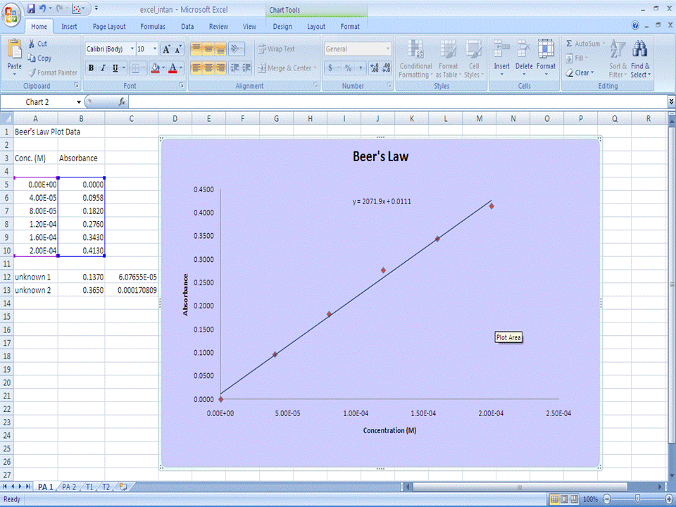
 Part 1 - Beer's Law Scatter Plot and Linear Regression


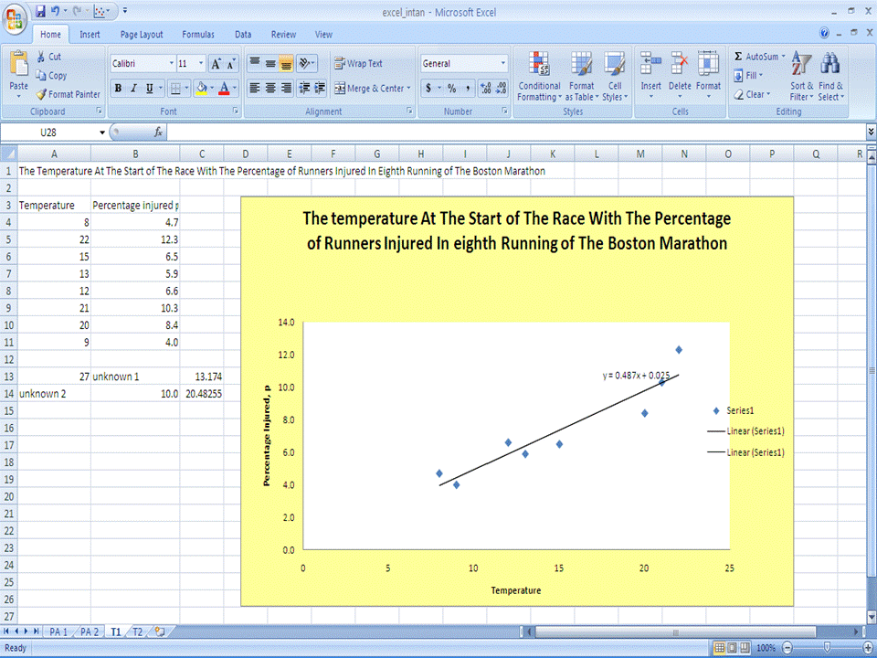
 Part 2 - Titration Data Plotting


  • Finding the line of best fit


  • Quadratic regression


Monday, January 3, 2011

CHEMSKETCH

Energy of Reaction Diagram


p-orbital, d-orbital, pi-type orbital


Vacuum Distillation Apparatus


Two-chain DNA Strand


Lipid

HTML


HTML?. What is HTML actually?.Many people did not really knew the exact meaning of HTML.
HTML  is a markup language used to structure text and multimedia documents and to set up hypertext links between documents, used extensively on the World Wide Web.

Read more: http://www.answers.com/topic/html#ixzz18dLW1nMq

HTML, which stands for HyperText Markup Language, is the predominant markup language for web pages.
A markup language is a set of markup tags, and HTML uses markup tags to describe web pages.

HTML is written in the form of HTML elements consisting of "tags" surrounded by angle brackets (like <html>) within the web page content. HTML tags normally come in pairs like <b> and </b>. The first tag in a pair is the start tags, the second tag is the end tags (they are also called opening tags and closing tags).

The purpose of a web browser is to read HTML documents and display them as web pages. The browser does not display the HTML tags, but uses the tags to interpret the content of the page.

HTML elements form the building blocks of all websites. HTML allows images and objects to be embedded and can be used to create interactive forms. It provides a means to create structured documents by denoting structural semantics for text such as headings, paragraphs, lists, links, quotes and other items. It can embed scripts in languages such as JavaScripts which affect the behavior of HTML webpages.

HTML can also be used to include Cascading Style Sheets (CSS) to define the appearance and layout of text and other material. The W3C, maintainer of both HTML and CSS standards, encourages the use of CSS over explicit presentational markup.

With the dynamic HTML color codes chart you can get codes for basic colors. See the chart and table below:

HTML COLOUR CHART


COLOURHTML CODE
blue#0000FF
Black#000000
Red#FF0000
White#FFFFFF
Green#008000
Purple#800080
Yellow#FFFF00
Orange#FFA500
Violet#EE82EE
Silver#C0C0C0
Gold#FFD700
Gray#808080
Pink#FFCOCB
Fuscia#FF00FF
light blue#ADD8E6
Sky blue#87CEEB
Aqua#00FFFF
Khaki#F0E68C


PROTEIN DATA BANK

INTRODUCTION

The PDB archive contains information about experimentally-determined structures of proteins, nucleic acids, and complex assemblies. As a member of the wwPDB, the RCSB PDB curates and annotates PDB data according to agreed upon standards.
The RCSB PDB also provides a variety of tools and resources. Users can perform simple and advanced searches based on annotations relating to sequence, structure and function. These molecules are visualized, downloaded, and analyzed by users who range from students to specialized scientists. 
The  Protein Data Bank (PDB) is a repository for the 3-D structural data of large biological molecules, such as proteins and nucleic acids. (See also crystallographic database). The data, typically obtained by X-ray crystallography or NMR spectroscopy and submitted by biologists and biochemists from around the world, are freely accessible on the Internet via the websites of its member organisations (PDBe, PDBj, and RCSB). The PDB is overseen by an organization called the Worldwide Protein Data Bank, wwPDB.
The PDB is a key resource in areas of structural biology, such as structural genomics. Most major scientific journals, and some funding agencies, such as the NIH in the USA, now require scientists to submit their structure data to the PDB. If the contents of the PDB are thought of as primary data, then there are hundreds of derived (i.e., secondary) databases that categorize the data differently. For example, both SCOP and CATH categorize structures according to type of structure and assumed evolutionary relations; GO categorize structures based on genes.

STRUCTURE

HtrA

LonA
ClpP
SUMMARY

For HtrA, (Rv3671c) a putative serine protease, is crucial for persistence of Mycobacterium tuberculosis in the hostile environment of the phagosome. We show that Rv3671c is required for M. tuberculosis resistance to oxidative stress in addition to its role in protection from acidification. Structural and biochemical analyses demonstrate that the periplasmic domain of Rv3671c is a functional serine protease of the chymotrypsin family and, remarkably, that its activity increases on oxidation. High-resolution crystal structures of this protease in an active strained state and in an inactive relaxed state reveal that a solvent-exposed disulfide bond controls the protease activity by constraining two distant regions of Rv3671c and stabilizing it in the catalytically active conformation. In vitro biochemical studies confirm that activation of the protease in an oxidative environment is dependent on this reversible disulfide bond. These results suggest that the disulfide bond modulates activity of Rv3671c depending on the oxidative environment in vivo. 


 For Lon ATP-dependent proteases are key components of the protein quality control systems of bacterial cells and eukaryotic organelles. Eubacterial Lon proteases contain an N-terminal domain, an ATPase domain, and a protease domain, all in one polypeptide chain. The N-terminal domain is thought to be involved in substrate recognition, the ATPase domain in substrate unfolding and translocation into the protease chamber, and the protease domain in the hydrolysis of polypeptides into small peptide fragments. Like other AAA+ ATPases and self-compartmentalising proteases, Lon functions as an oligomeric complex, although the subunit stoichiometry is currently unclear. Here, we present crystal structures of truncated versions of Lon protease from Bacillus subtilis (BsLon), which reveal previously unknown architectural features of Lon complexes. Our analytical ultracentrifugation and electron microscopy show different oligomerisation of Lon proteases from two different bacterial species, Aquifex aeolicus and B. subtilis. The structure of BsLon-AP shows a hexameric complex consisting of a small part of the N-terminal domain, the ATPase, and protease domains. The structure shows the approximate arrangement of the three functional domains of Lon. It also reveals a resemblance between the architecture of Lon proteases and the bacterial proteasome-like protease HslUV. Our second structure, BsLon-N, represents the first 209 amino acids of the N-terminal domain of BsLon and consists of a globular domain, similar in structure to the E. coli Lon N-terminal domain, and an additional four-helix bundle, which is part of a predicted coiled-coil region. An unexpected dimeric interaction between BsLon-N monomers reveals the possibility that Lon complexes may be stabilised by coiled-coil interactions between neighbouring N-terminal domains. Together, BsLon-N and BsLon-AP are 36 amino acids short of offering a complete picture of a full-length Lon protease.

We have determined the crystal structure of the proteolytic component of the caseinolytic Clp protease (ClpP) from E. coli at 2.3 A resolution using an ab initio phasing procedure that exploits the internal 14-fold symmetry of the oligomer. The structure of a ClpP monomer has a distinct fold that defines a fifth structural family of serine proteases but a conserved catalytic apparatus. The active protease resembles a hollow, solid-walled cylinder composed of two 7-fold symmetric rings stacked back-to-back. Its 14 proteolytic active sites are located within a central, roughly spherical chamber approximately 51 A in diameter. Access to the proteolytic chamber is controlled by two axial pores, each having a minimum diameter of approximately 10 A. From the structural features of ClpP, we suggest a model for its action in degrading proteins.